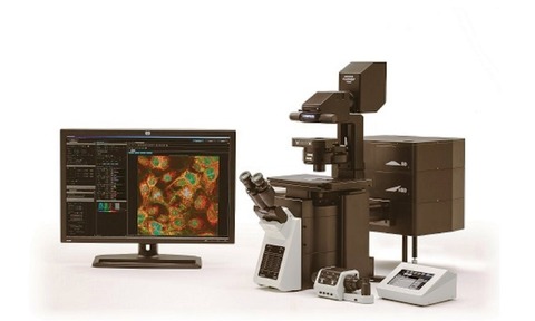
Olympus’ FLUOVIEW FV3000 confocal laser scanning microscope combines high-performance imaging capabilities with ease of use so that researchers can collect relevant imaging data quickly and easily.
Built for fast, stable and accurate measurements of biological reactions within living cells and tissues, Olympus’ FV3000 offers flexibility for all live-cell imaging applications, providing high-resolution images of structures and dynamic intracellular processes.
Succeeding the FV1200, the FV3000 is controlled via an intuitive software interface.
Its optical design offers macro to micro imaging capabilities with objectives ranging from 1.25X to 150X magnification. The FV3000 features a galvanometer scanner
whilst the FV3000RS offers a hybrid resonant/galvanometer scanner.
The high-speed resonant scanner can acquire high-resolution images at up to 438 frames per second.
The Olympus TruSpectral detection system is optimised for maximum efficiency when delivering emission light to detectors. Users can select the exact wavelength range they want to capture in every detection channel at nanometer precision.
FLUOVIEW software has been designed with an intuitive interface that can be tailored to adapt to the way an individual works. Workflows can be created and saved, and the interface customised for easy access to the most commonly used applications.


