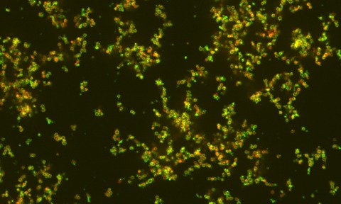
Using SEEC microscopy, French scientists have discovered that bacterium adhesion and motion are driven by the same mechanism.
A scientific endeavour carried out by two French groups belonging to INSERM and CNRS at Aix-Marseilles University shows for the very first time that both bacterium adhesion to and bacterium motion on a surface are driven by the same mechanism.
Those findings result from collaborative work with Nanolane, a French company that specialises in optical characterisation. Nanolane has devised a new generation of advanced microscope slides specifically for this type of investigation.
Up until now, it was believed that the motion of a bacterium on a surface was caused by projection of a polymeric material referred to as slime that would be produced at the bacterium rear.
The Marseilles-based scientists’ work, recently published in America’s Proceedings of the National Academy of Sciences, proves that slime is being generated at spots spread out all along a bacterium’s body as opposed to the rear as seemed to be agreed upon in the scientific literature.
The role played by this slime appears to be two-fold: it works as a glue so that a bacterium can stick to a surface, and it facilitates bacterium motion by lubricating the surface-to-bacterium contact.
It is because of the picture quality SEEC microscopy provides by substituting Surf slides for ordinary microscope slides that the conclusions were drawn.
Surfs are the new generation of microscope slides that Nanolane developed, they have the power to let a reflected light microscope image samples in an aqueous medium with a sensitivity level of under one nanometre.


