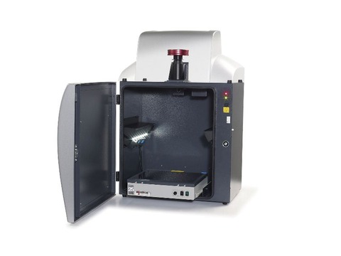
Cambridge University is using the G:BOX XR5 to understand which genes are involved in remyelination.
The G:BOX XR5 imaging system is being used at the University of Cambridge to visualise and analyse DNA as part of a research programme to understand the molecular mechanisms behind why the remyelination process fails in diseases such as Multiple Sclerosis (MS).
Scientists in the Neurosciences Group at the Department of Veterinary Medicine, part of the University of Cambridge are using a G:BOX XR5 system to accurately image gels of fluorescent dye stained PCR products derived from genes involved in the remyelination process.
By studying these genes, the scientists hope to find a means of enhancing the repair of normally non-repairable clinical conditions such as MS.
Dr Chao Zhao Assistant Director of Research, in the Neurosciences Group said: “Our research focuses on the mechanism of remyelination of the central nervous system after demyelination. To do this we are trying to understand the molecular mechanism of why myelin repair fails in diseases such as MS and we use the G:BOX XR5 every day to visualise and sometimes analyse very small amounts of PCR products as part of this research.”


