Scientists discover structure of protein essential for quality control
15 Jan 2013

The Scripps Research Institute (TSRI) has pushed the boundaries of electron microscopy to determine the structure of Ltn1.
Ltn1 appears to be essential for keeping cells’ protein-making machinery working smoothly. It may also be relevant to human neurodegenerative diseases, for an Ltn1 mutation in mice leads to a motor-neuron disease.
“To better understand Ltn1’s mechanism of action, we needed to solve its structure, and that’s what we’ve done here,” said TSRI Associate Professor Claudio Joazeiro.
“In addition, this project has brought us a set of structural analysis techniques that we can apply to other exciting problems in biology,” said TSRI Professor Bridget Carragher.
Links to neurodegenerative disease
Ltn1 first turned up on biologists’ radar screens several years ago when a joint Novartis-Phenomix research team noted that mice with an unknown gene mutation were born normal but suffered from progressive paralysis.
In a study published in the journal Nature the following year, Joazeiro and his postdoctoral research associate Mario H. Bengtson found that the enzyme serves as a crucial quality-control manager for the cellular protein-making factories called ribosomes.
Occasionally a ribosome receives miscoded genetic instructions and produces certain types of abnormal proteins, known as “nonstop proteins” – jamming the ribosomal machinery like a wrinkled sheet of paper in an office printer.
Bengtson and Joazeiro found that Ltn1 fixes jammed ribosomes by tagging nonstop proteins with ubiquitin molecules, thereby marking them for quick destruction by roving cellular garbage-disposers called proteasomes.
“The question for us then was, “How does Ltn1 do this?’ ” said Joazeiro.
Pushing the boundaries of electron microscopy
To help find out, he began a collaboration with Carragher and Potter, who run the National Resource for Automated Molecular Microscopy (NRAMM), an advanced electron microscope facility at TSRI that is funded by the National Institutes of Health’s National Center for Research Resources.
No one has ever used electron microscopy to distinguish so many conformations of such a small protein
Ltn1 was deemed too large for its structure to be determined by current nuclear magnetic resonance (NMR) technology, and, as the scientists know now, too flexible to allow the highly regular crystalline packing needed by X-ray crystallographers. “It’s a very floppy molecule, so it would be hard to crystallize,” said Potter.
Advanced electron microscopy offered a way, however. Dmitry Lyumkis, a graduate student in the NRAMM laboratory and first author of the study, took high-resolution images of yeast Ltn1 with an electron microscope.
He then used sophisticated image and data processing software to align and average individual images. The technique eliminates much of the random “noise” that obscures single images and produces a sharp 3D picture of the protein.
No one has ever used electron microscopy to distinguish so many – more than 20 – conformations of such a small protein.
“Usually electron microscopists determine no more than two or three conformational states, and they work with protein complexes whose size is in the megadalton range, but Ltn1 is only 180 kilodaltons, an order of magnitude smaller,” Lyumkis said.
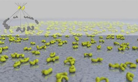
The analysis revealed that Ltn1 has an elongated, double-jointed and extraordinarily flexible structure with two working ends – the N-terminus and C-terminus.
“We anticipate that the N-terminus is responsible for association with the ribosome and know that the C-terminus is responsible for the ubiquitylation of nonstop proteins,” said Lyumkis. “We suspect that the high flexibility of this structure is needed for it to work on the variety of nonstop proteins that can get stuck in ribosomes.”

