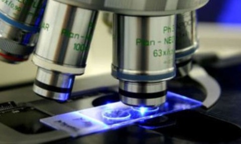
Microscopy techniques to benefit from a £25.5m funding
27 Feb 2013

Biomedical research in the UK is to benefit from a £25.5m cash injection to advance microscopy techniques.
Three of the UK’s research councils have invested £25.5m to establish 17 microscopy platforms that are hoped to bring about advances in biological and biomedical research.
The funding was awarded by the Medical Research Council, the Biotechnology and Biological Sciences Research Council and the Engineering and Physical Sciences Research Council.
Recent advances in microscopy builds dramatically on the previous limits of electron and light (optical) microscopy. Electron microscopy has very high resolution but can not be used to image living cells or organisms. Traditional light microscopy can look at living materials but has far lower resolution.
This type of microscopy relies on scientists in very different disciplines coming together
The new generation of imaging techniques are now able to greatly increase the resolution - sometimes to close to molecular level - when studying an intact and living cell.
These structures are some of the smallest things that scientists have been able to visualise. For example, a cell membrane is about 6-10 nanometres (a nanometre is 1 millionth of a millimetre).
As well as increasing the magnification, researchers are now able to study live biological processes as they are taking place at fractions of a second.
Being able to visualise these tiny biological structures, such as the proteins involved in cell function and the biological and chemical processes in which they are involved, will allow researchers to understand more about what causes disease.
Professor Steve Hill, who chaired the expert panel which assessed the proposals, said: “Microscopy is one of the most important tools scientists have for discovery-based research but the high costs associated with this technology are often a barrier to expansion.
“This type of microscopy relies on scientists in very different disciplines coming together to solve very specific imaging problems.
“All seventeen projects were able to demonstrate extremely strong partnerships between biologists, physicists, chemists, mathematicians, engineers, technologists and equipment manufacturers.”
Awards included:
- Scientists at the University of York are combining both electron and light microscopy techniques. One application will be to find out more about neurodegenerative disease by visualising the processes in a nerve cell which govern learning and memory.
- A partnership between biological researchers at the MRC Clinical Sciences Centre and physicists at Imperial College London to visualise fundamental biological mechanisms and benefit on-going research in areas such as the behaviour of chromosomes in developing reproductive cells, the function of nerve cells and the regulation of gene expression.
- At the University of Leeds, researchers will be building a new microscope to observe the internal structure of cells and gain insights into the development of conditions such as cancer and cardiovascular disease.

