Atom-by-atom imaging technique revealed
11 Jun 2013

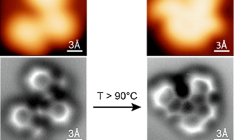
Researchers from the University of California have developed a technique that captures atomic scale reactions of a chemical.
The scientists used an atomic force microscope (AFM) to capture how an atoms molecular structure changed during a reaction.
Until recently, scientists had only been able to assume this type of information from spectroscopic analysis.
Felix Fisher, lead researcher on the project, stated: “Even though I use these molecules on a day to day basis, this was what my teachers used to say that you would never be able to actually see, and now we have it here.”
It is believed that the research conducted will enable chemistry students to better understand molecular reactions and allow them to fine-tune their reactions to create the products they want.
The basis for Fisher’s research was to capture the images in the hope that he and his team could build innovative graphene nanostructures for use in next-generation computers.
This was what my teachers used to say that you would never be able to actually see, and now we have it here
“The implications go far beyond just graphene,” stated Fisher. “This technique will find application in the study of heterogeneous catalysis, for example.”
Fisher argued that to understand the chemistry that is actually happening on a catalytic surface, we need a tool that is very selective.
Michael Crommie, a research partner on the team, believes that: “The atomic force microscope gives us new information about the chemical bond, which is incredibly useful for understanding how different molecular structures connect.”
Crommie suggested that the information gained is essential for creating new nanostructures such as bonded networks of atoms that have particular shapes and structures.
The original technique developed by Fisher sought to construct graphene nanostructures that possessed unusual quantum properties, designed for nano-scale electronic devices.
Fisher and Crommie then devised a way to chill the reaction surface and molecules of the atomic structures to the temperature of liquid helium - almost 4 Kelvin.
This technique stopped the molecules from moving around too erratically so that a scanning tunnelling microscope could locate the molecules on the surface of the graphene, which is then probed more finely with an AFM.
To enhance the spatial resolution, the team added a carbon monoxide molecule to the tip, a technique known as non-contact AFM.
“Ultimately, we are trying to develop new surface chemistry that allows us to build higher ordered architectures on surfaces, and these might lead into applications such as building electronic devices, data storage devices or logic gates out of carbon materials,” concluded Fisher.

