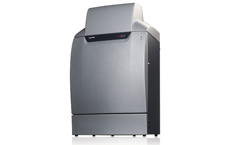
Maastricht University is using a Syngene G:BOX chemi imaging system as part of its science bachelors programme.
Students at the Chemelot Campus are using a G:BOX chemi system to accurately image proteins extracted from mosses, tropical leaves and insects run on Coomassie blue stained gels or blotted onto chemiluminescent Western blots. This approach is allowing the students to quickly and easily analyse a range of proteins seen in the natural environment.
Paul Lemmens, lab coordinator, Maastricht Science Programme said: “Our Science Bachelors Programme allows our students to undertake plenty of practical laboratory work. Being able to understand and perform molecular biology techniques is crucial for many scientists today, and we wanted an imaging system that would allow our students to easily switch applications, to train them to analyse a range of different gel and blot types.”
“We assessed two imaging systems but felt that the Syngene system’s software was much easier for the students to use with minimal training. From using the G:BOX Chemi we have found the quality of the apparatus is very good and our enthusiastic students can now use the system without any difficulty,” he added.
Laura Sullivan, SDI’s Syngene divisional manager commented: “At Syngene we realise that not everyone using image analysis is an expert at understanding how cameras work to image gels and blots. This is why we developed our GeneSys software to make it easy for a novice to set the conditions up to get perfect images of their research with our G:BOX systems.
“We’re delighted that Maastricht University has seen the benefits of using this software and their use of the system shows how well suited a G:BOX Chemi image analyser is for any university molecular biology programme to use in training the next generation of high calibre life scientists,” concluded Sullivan.


