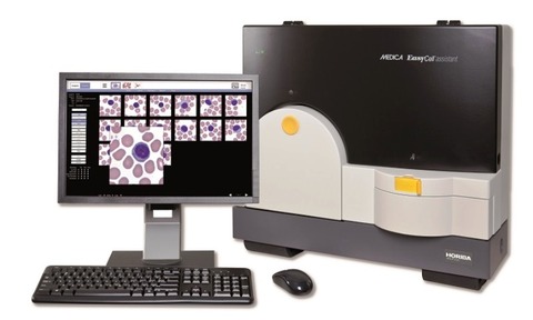
The imaging system can improve laboratory productivity by automatically scanning and locating up to 200 white blood cells on a smear slide and pre-classifying them on a display, grouped by cell type.
The EasyCell assistant digital cell imaging system from Horiba Medical is designed to simplify manual differentials by pre-classifying normal cells, enabling rapid validation and freeing time to devote to the review of abnormal cells which are displayed separately.
Red blood cells and platelets are also displayed for morphology comments and platelet estimates provided to verify the Full Blood Count (FBC) result.
The EasyCell assistant also saves time by enabling walk-away operation, since slides can be loaded into its 30-position carousel and left to process.
However, when immediate analysis is required they can be placed in the EasyCell Stat position. All slides can also be directly accessed during the scanning process should the operator wish to review them immediately.
The system can also handle a variety of stained slides, including Wright, Wright-Giemsa, or May-Grünwald-Giemsa stains.
For greater efficiency and verification, all scanned slide images can be securely shared between multiple users remotely by installing EasyCell Remote software on existing computers within the hospital network and even via VPN.
EasyCell Remote workstations hold all the functionality of an EasyCell assistant, except for slide processing. This enables ready viewing for competence training and monitoring lab quality by managers, or convenient referral for second opinions on difficult cases.
This also makes it an ideal teaching tool since detailed, magnified cell images can be simultaneously viewed by instructor and student.
The system is highly accurate with unique, on board quality control software that verifies the consistent and correct location and presentation of nucleated cells.
The QC programme also checks for acceptable slide preparation and system hardware operation.
Furthermore, since the system automates data access, all slide images can be archived with accompanying reports of patient demographics, results and comments, enabling full traceability and rapid access at a later date to track patient status over time.


