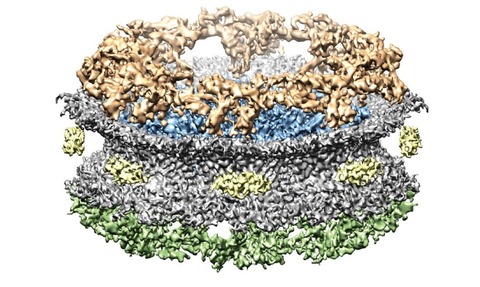
High-tech microscope displays nuclear pores
26 Jun 2015

Researchers at the University of Zurich have used cryo-electron microscopes to display the pores in the cell nucleus with greater precision.
As a result of the study, the Swiss research team discovered a previously unobserved structure inside the nuclear pore that forms a kind of molecular gate.
Lead researcher Ohad Medalia said the gate can only be opened by molecules that “hold the right key”.
“The gate can only be opened by molecules that hold the right key
Lead researcher Ohad Medalia
According to Medalia, the molecular gate is the so-called spoke ring, which is sandwiched between two other rings and extends inside the nuclear pores. The gate itself consists of a fine lattice, which enables small molecules to slip through unobstructed.
Complex structures
In a human cell, more than a million molecules are transported into the cell nucleus every minute. In the process, pores embedded in the nucleus membrane act as transport gates - as outlined in the university’s research.
These nuclear pores are considered to be among the largest and most complex structures in the cell and comprise more than 200 individual proteins, which are arranged in a ring-like architecture - containing a transport channel.
Medalia’s team has, for the first time, succeeded in displaying the spatial structure of the transport channel in the nuclear pores in high resolution.
The breakthrough could lead to a better understanding of why certain molecules are allowed to pass through the nuclear pores while others are turned away, the researchers claimed.
It may also help improve our understanding of the development of some diseases that involve a defective transportation to the nuclear pores - such as intestinal, ovarian and thyroid cancer.

