Viral vision: intravital microscopy
13 Jul 2015

A research lab in Italy is using intravital microscopy to study virus responses.
Matteo Iannacone, a group leader in the Division of Immunology at the San Raffaele Scientific Institute, Milan is attempting to dissect the complex dynamics of host-virus interactions - paying particular attention to the development and function of adaptive immune responses.
But as it is still beyond the reach of even the most sophisticated in vitro methodology to simulate the complex interplay of physical, cellular, biochemical and other factors that influence cell behaviour in microvessels and interstitial tissues, the Milan-based group uses intravital microscopy.
This technique is complemented by more traditional molecular, cellular and histological approaches, thereby characterising host-virus interactions at the molecular, single cell and whole animal level.
Two-photon microscopy
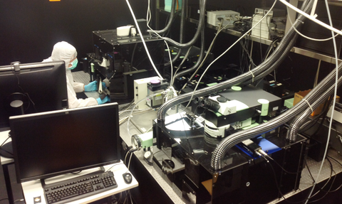
Specifically, Iannacone’s group is using a TriM Scope II 2-Photon microscope designed by LaVision BioTech - a German imaging manufacturer that develops advanced microscopy solutions for life sciences applications.
TriM Scope II is a modular multi-photon/confocal microscopy platform that combines single-beam and multi-beam operation in one microscope.
This allows for deep in-vivo imaging, LaVision says.
“The choice of the TriM Scope from LaVision was driven by the desire of the group to couple a state-of-the-art multiphoton microscope with the possibility of customising the entire system to our need to work in vivo,” Iannacone says.
“In these studies, both confocal immunofluorescence histology and correlative light-electron microscopy are used too,” he adds.
Blood flow velocity
In vivo microscopy makes it possible to map blood flow velocity at a high spatial and time resolution in the complex network of vessels constituting the hepatic microcirculation in the liver, while simultaneously maintaining the morphologic information related to the dynamic parameters of vessels and immune cells.
Iannacone’s ongoing project with the Department of Physics at the University of Milano-Bicocca was recently reported online via the journal Cellular & Molecular Immunology.
The article considers the potential of various novel imaging techniques to show how CD8+ T-cells move through the liver and how this may impact on the pathogenesis of Hepatitis B Virus, HBV.

