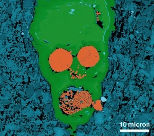
Jeol has used one of its scanning electron microscopes to study the potential of oil shale.
Abundant in specific regions of the US, oil shale is a fine-grained, sedimentary rock composed of flakes of clay minerals and tiny fragments of other minerals, especially quartz and calcite.
Shale also has a complex network of soft veins of an organic substance, kerogen and accessory opaque minerals such as pyrite.
When heated, kerogen can release hydrocarbons, or fossil fuel.
By studying the internal composition of the shale and the network of kerogen-filled veins, scientists can determine the abundance and ease of extraction of oil.
Companies investigating this alternative source of energy have turned to Jeol for not only the electron microscope, but also a sample preparation tool that creates pristine cross sections of oil shale.
Without this tool, it is difficult to prepare cross sections of shale without distorting and smearing the soft veins of kerogen trapped in the composite.
The Jeol cross-section polisher sliced and polished the sliver of shale with an argon beam to yield undistorted, precise cross sections.
Every detail can be clearly seen, allowing researchers to clearly see the network of veins of kerogen in the sample, and can take the imaging a step further to make 3D reconstructions of the pore network by using a serial slicing and sampling technique with the multibeam focused ion beam (FIB) instrument.


