Amsbio has announced a new range of ready-to-use lentiviral particles that allow direct visualisation of organelles or structures in cells without manipulation
For real time visualisation, traditionally researchers have had to use plasmids for organelle markers, but the labelling was transitory.
Now using the new lentiviral particles, Amsbio claims researchers have an easy way to provide long-term labels on many cellular structures (stable integration inside the genome) without the need for transfection reagents.
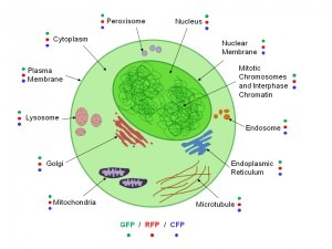
Using lentiviral particles also allows effective direct visualisation of Nuclei, Nuclear Membrane, Mitotic Chromosomes and Interphase Chromatin, Endosome, Endoplasmic Reticulum, Microtubule, Mitochondria, Golgi, Lysosome, Plasma Membrane, Cytoplasm and Peroxisome in hard to transfect mammalian cells, stem cells and primary cells.
The new lentiviral particles employ three fluorescent protein tags (GFP, RFP or CFP), fused to specific DNAs encoding organelle-specific or structure-specific proteins, to enable direct visualisation of the organelles or structures without manipulation or antibodies.


