The right angle
27 Feb 2013
Ceram’s Giles Blundred discusses how to push the boundaries of X-ray diffraction beyond routine 2 theta measurements.
X-ray diffraction (XRD) is a mature characterisation technique for materials characterisation.
Typically, this technique delivers quantification of the crystalline phases present for a given solid with additional information concerning the average unit cell size for a given phase, the average crystallite size and the average residual strain for any given phase present.
Applications range from quantitative Phase Analysis of cements to crystallite size determination of silver halides used in photographic film.
Critically for such characterisation, an X-ray diffractometer is set up to run in Bragg-Brentano para-focusing mode. This means there is a divergent x-ray beam that is “reflected” through a sample to be focused on the detector.
In this mode, where the incident beam angle and the detector angle vary symmetrically about the normal to the sample surface with increasing two theta, the X-ray penetration depth increases with increasing incident angle.
During the late 1990’s the collimation of X-ray beams using graded synthetic crystals moved forward with the onset of the Goebel mirror. This optic when placed into an x-ray beam allows a divergent beam to be collimated to a parallel beam.
The use of a parallel beam optic allows X-ray Diffraction to be used in situations that were previously accessible to specialised machines. Namely, the thickness and roughness of surface layer coatings placed at the surface of a sample, akin to passivating oxide layers on metals or corrosion products at the surface of a sample.
In order to gather data related to the surface of fabricated materials Grazing Incidence of Diffraction must be employed. The fact that the incident beam impinges upon the sample surface at a fixed incident angle allows for controlled beam penetration depths within a given sample.
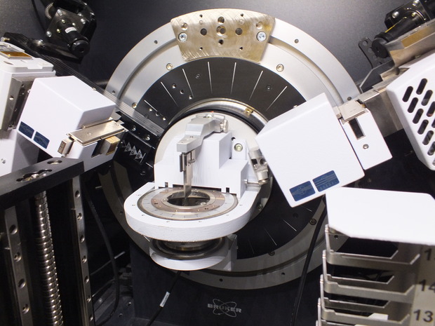
This technique is most suitable for thin film layers deposited on very flat surfaces. A typical example is afforded using the sputtering of a thin gold film onto a glass substrate. GID traces were obtained using a Bruker D8 Advance.
Copper K alpha radiation was employed and the incident beam was collimated through a Goebel Mirror and a 0.2mm slit on the incident side. In order to maintain a parallel beam geometry a long equatorial soller slit was used prior to the Lynxeye solid state detector used in 0d mode. Incident angles used were 0.2, 0.5, 0.6 0.75 and 1.0.
It can be seen with increasing incident angle there is an increasing signal from amorphous glass slide substrate with a diffuse scattering peak at 23? two theta. The estimated thickness of the gold layer is 20 nanometres. X-Ray Reflectometry on the same sample indicates that the thickness of the sputtered gold layer is 12 nanometres.
Evaluation of the presence of crystalline gold at the surface and the thickness of the gold layer is only afforded by the use of GID. If normal Bragg-Brentano geometry XRD was used then the XRD trace would have been dominated by the amorphous glass substrate below the gold coating and the thickness of the coating could not be determined.
The Grazing Incidence of diffraction technique is used to qualitatively evaluate the formation of Hydroxy-apatite-like precipitates in the international standard BS ISO 23317:2007 “Implants for surgery - In vitro evaluation for apatite-forming ability of implant materials”.
Bio
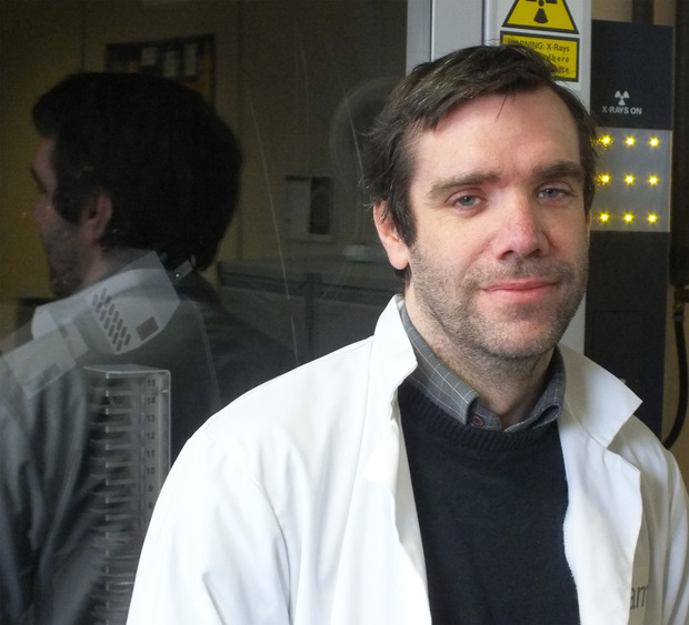
Giles Blundred, X-ray diffraction scientist at Ceram
Giles graduated with dual honors in Chemistry and Geology from Keele University UK. His PhD thesis was based on the characterisation of Mixed Ionic and Electronic Conducting Oxides by correlating the observed physical properties with the structural properties. Giles has over 11 years’ experience of using diffraction techniques in the characterisation of the structure of solids.


