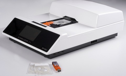
Denator AB’s heat-stabilisation system has been shown to be compatible with MALDI imaging analysis.
The preservation of sample components prior to MALDI imaging analysis is crucial to accurately measure the distribution and abundance of biological molecules in organs.
During downstream analysis, the presence of metabolites, drug compounds and endogenous peptides, existing at very low levels in the tissue sample, is often difficult to detect due to rapid degradation of the molecule of interest or masking by protein fragments generated from normal degradation processes.
MALDI was used alongside Denator’s Stabilisor system - based on the company’s heat-stabilisation technology - to stop degradation from the moment of sampling.
This leads to increased accuracy and quality of analytical results. Denator says that such utilisation will be of particular interest to researchers within pharmaceutical research looking at mapping molecules in situ on tissue sections.
A recently published paper demonstrated an increased recovery of metabolites from tissue samples when using heat stabilisation followed by an optimal protocol for MALDI imaging analysis (Conductive carbon tape used for support and mounting of both whole animal and fragile heat-treated tissue sections for MALDI MS imaging and quantification, Goodwin et al., Journal of Proteomics, 2012).
Olof Sköld, CEO at Denator, says: “MALDI imaging is a very promising tool for biomarker characterisation and localisation in drug development.
“By working closely with users of our system, we have now proven the compatibility between our Stabilisor system and this analytical method. We see this development as a natural step to ensure that researchers who heat-stabilise tissues upstream achieve the best possible analytical results downstream.”


