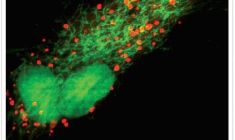
The secret life of cells
6 Jul 2015

Instruments that can capture the covert processes within cells are bringing laboratory research into much sharper focus, writes Louisa Hearn.
The imaging of live cells has ushered in a brave new era for researchers tasked with observing and recording delicate biological processes as they naturally unfold.
A new generation of sophisticated devices are easing the journey for the rising number of researchers choosing live cell imaging over the more traditional method of fixed cell imaging, say experts.
“Because you are trying to work out what is happening in a biological process, the closer you can get to real life the better
Carl Zeiss imaging specialist Chris Power
The most important advantage that live cell imaging offers over fixed cell imaging is that it is inherently more trustable, says Chris Power, 3D imaging specialist at imaging systems firm Carl Zeiss.
“Because you are trying to work out what is happening in a biological process, the closer you can get to real life the better.”
A fixed cell sample, which is still the most common form of cell imaging technology in use, is literally fixed in place with the use of chemicals so it can be viewed in a very high resolution.
Live cell imaging, on the other hand, requires an optimum environment to preserve the health of the cells for the duration of the experiment.
You can also acquire far more data from a live environment, while using fewer samples, Power says.
This is particularly important amid tightening the regulatory standards for conducting animal research.
“In the past, if scientists had been studying a particular genetic abnormality in, for example, a fish, and they wanted to track how its heart developed, they would have had to study hundreds of fish to attain that data,” he says.
“Now they can take just one organism and image it for the entire development cycle of a biological process. Not only is this more reliable but only a single fish would be required for the research.”
Because they want their sample to be as viable at the end of this process as it was at the start, the fish would most likely be able swim away at the end of the experiment, says Power.
The largest sector of the live cell imaging market for Carl Zeiss comprises instruments designed for optical sectioning of samples, says Power.
The foundation of this technology was the confocal microscope, which came to market in 1984, and since then many others have followed.
Used almost exclusively in university laboratories, what sets different types of instruments apart is “speed, sensitivity, and resolution”, he says.
“Now there are many different ways of optical sectioning, and the type of microscope you might use depends on whether you are looking at individual cells or an entire mouse brain.”
Organogenesis
From live cell imaging two new exciting avenues of study have emerged, says Power.
One of these is Organogenesis, which is the creation of organs and cell lineage.
“So if you want to know where the cells in eyes came from, within that initial ball of cells in a developing organism, you just image the process all the way to the development of the eye and then play the movie backwards.”
The emerging field of Optogenetics takes this process a step further by introducing cells that have been genetically modified with fluorescence.
“You can engineer some of these cells to behave differently if you shine certain wavelengths of light on them, so while you are shining the light, you can identify the function of specific cells in your sample as they are lit up.”
These types of high-end research projects require multidisciplinary teams, says Power.
These might include biologists, biochemists as well as bioinformatics and programmers to manage and interpret the massive volume of data they generate.
This also has ramifications for those looking to publish research findings.
“A few years ago you could publish on very little data, but now there is very much a trend to do it on a good sample size”, says Power.
“This means we are moving to far more automated processes within microscopes that will automatically acquire images of hundreds of cells.”
When it comes to published research, the quality of image or movie that is captured can also have a big impact on the level of exposure it can generate.
“Fixed cell imaging will allow you to create statistical data, and it may also give you the cover shot,” says Magnus Persmark, a senior product manager of cellular analysis at Thermo Fisher Scientific.
“However, to capture the dynamics of cellular processes like membrane trafficking, differentiation or cell migration, the ability to image live cells is essential; the data represented in a time-lapse movie of cells dividing or doing something else over time is both beautiful and informative.”
When you are involved with tissue engineering, three-dimensional co-cultures or other complex models, working with living substrates is a requirement, adds Persmark.
The cellular analysis unit of Thermo Fisher Scientific has for several years focused efforts on developing instruments that are relatively easy to use, but still give the requisite image and data quality, he says.
“We offer a full complement of instruments for analysis of cells by microscopy, flow cytometry or cell counting, in particular using fluorescence-based assays,” says Persmark.
“They might be used for something as pedestrian as looking at cell confluence or number, whether they are alive or dead, or taking a sample from a bioreactor to see if the cells express Green Fluorescent Protein (GFP),” he says.
“General areas of cell-based research where we see a lot of activity and interest are biomedical applications such as oncology and neurology, as well as research on age-related conditions such as diabetes and Alzheimer’s.”
“There is a range of cell models for which we have developed reagents for fluorescence-based assays or procedures. These might be mammalian, microbial yeast or bacteria, but they all involve cells in some shape or form.”
’Significant developments’
Until recently fixed cell imaging accounted for 70% of all cell based fluorescent assays, says Persmark.
However, this is changing rapidly owing to “incredible developments in cell and molecular biology as well as fluorescent reagents.”
These have sprung from the discovery of GFP “which has really afforded us an ability to label cells and cellular components,” he says.
There have also been significant developments in instrumentation, he says.
“On the very cutting edge of research are new modalities in very high resolution imaging that enable visualisation of structures right down to the refraction limit of light, enabling us to probe even deeper into cellular details,” he says.
“Today, instruments also exist that allow imaging of live cells over several days, which in conjunction with advanced software algorithms provide visual and quantitative data unimaginable just a few years ago.”

