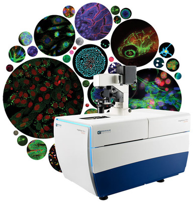
With growth of new 3D cell culturing techniques and even whole organism models, image acquisition and analysis of such systems is becoming increasingly important.
Molecular Devices have published an ebook about 3D Cell Imaging and Analysis which outlines some of the 3D cell model applications and approaches for acquiring and visualising quantitative data.
Now that highly complex 3D cellular models, such as cancer spheroids and stem cell matrixes, are available, which better simulate the in vivo environment than 2D cultures, quantitative assays using such cultures have become a very attractive proposition, however image acquisition and analysis of such 3D models has proven challenging.
Next-generation, high-content, high-throughput microscopy tools, such as the ImageXpress Micro Confocal High-Content Imaging System and MetaXpress 3D Analysis Software from Molecular Devices, now offer innovative and automated techniques for evaluating the complex biology of these advanced models. In addition to 3D models, the technology is also applicable to whole organisms, such as zebrafish, as well as a wide variety of subcellular assays.
This eBook highlights some of these applications as well as Molecular Devices solutions for acquiring and visualising quantitative data. The ebook also provides useful links to further application notes, webinars, posters and articles about this rapidly evolving technique that is helping to span the gap between conventional 2D culturing and the in vivo environment. Get the ebook here.
To download the ebook click here.


