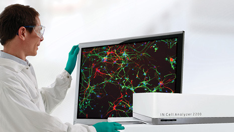
GE Healthcare application of automation to image acquisition and analysis is helping to take knowledge of cell structure and function to new levels of understanding.
In the C21st automation of both image acquisition and analysis has helped develop a more robust and productive approach to cell science.
Whether it is cancer research, cell biology, microbiology or drug delivery detailed examination of cells at the microscopic level enables you to probe a wider range of cell activities. From studying cell behaviour to elucidating molecular function, correlating multiple events and markers with cell morphology and unravelling complex signalling mechanisms, GE Healthcare are helping scientists to probe deeper and more efficiently.
The images in GE Healthcare’s latest promotional video illustrate just how they are helping to unlock greater detail about the basic structure and function of the cell using a number of different techniques.
High Content Analysis is a powerful technique for high through put microscopic analysis of cells. GE Healthcare’s IN Cell Analyser automated systems provide morphological and molecular data using high content cell imaging and analysis.
The combination of IN Cell Analyser HCA systems, IN Cell Investigator image analysis software, alongside IN Cell Miner data management software provides an integrated solution for high content cellular and sub cellular analysis in both basic and applied research. High and Super resolution Imaging provides advanced imaging solutions for cellular and subcellular visualisation.
GE Healthcare’s DeltaVision systems offer a range of imaging modalities and capabilities for most biological investigations. Advanced optical engineering and the latest technological developments in both high-resolution and super-resolution techniques combine in the DeltaVision Elite and DeltaVision OMX systems to offer highly flexible microscopy platforms supporting a wide variety of cell imaging needs, particularly live cell imaging.
Image Cytometry helps accelerate routine cellular analysis. GE Healthcare’s Cytell Cell Imaging System elegantly combines the functions of a digital microscope, a cell counter and an image cytometer in a single benchtop instrument. The Cytell Cell Imaging System provides a reliable and accurate method for researchers and technicians to easily capture cellular and subcellular image data and analysis results.
Cell-based assays can be optimised for more accurate representation of cell biology content. Cell-based tools are essential for studying cellular mechanisms in a biological context and a variety of cell- based assays using fluorescence technologies are available to cover many applications.
GE Healthcare produce a number of products for assessing cellular functions including the Cytell Cell viability Kit (and reagent version), Cytivia Cell Health Assay, Cytivia cell Integrity Assay and their Cell Proliferation Fluorescence Assay.
To see their high content analysis system in action please click here.


