Cells under the spotlight
30 Jul 2013

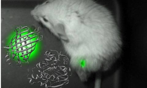
Experts at Osaka University have documented a luminescent protein which allowed real-time imaging of intracellular structures in living cells.
The theory of optogenetics enabled researchers to control the activity of individual neurons and measure the effects of those manipulations.
However, problems can arise when imaging fluorescence as the same light that stimulates the factors under optogenetic control can interfere with fluorescent sensors.
For example, the light which was used to animate the FRET-based indicator for calcium activated a commonly used photo-sensitive receptor used in optogenetics.
The technique is capable of viewing all biological phenomena
Osaka University professor Dr. Takeharu Nagai
University of Osaka professor Dr. Takeharu Nagai said: “Because fluorescent indicators require excitation light source, such excitation light gives perturbations to the cell function as photo-stimulation light.”
In order to overcome this problem, Dr. Nagai and his team developed a probe that fused a luminescent protein from the sea pansy Renilla reniformis with a fluorescent protein previously designed.
The protein, dubbed ’nano-lantern’, is believed to be the brightest in development and offered spatial and temporal resolution equivalent to those of fluorescence - according to the research team.
Upon completion of the novel protein, Nagai and his team set about testing its capabilities, imaging intracellular structures in living cells.
In collaboration with Dr. Yuriko Higuchi of Kyoto University, Dr. Nagai was able to use the nano-lantern to visualise cancer tissue within a mouse.
The technique allowed the research team to monitor and create video-rate imaging of tumors 17 days after implementation.
Subsequently, the team modified the protein into a calcium sensor and co-expressed it with a light-sensitive photoreceptor in rat neurons.
This co-expression system allowed the research team to follow excitation of the photoreceptors by measuring the calcium ion (Ca2+) increase as reported by the nano-lantern Ca2+ indicator.
In order to accurately visualise various nano-lantern signals the research team used a Photometrics’ Evolve 512 EMCCD camera.
The technology could be used for more advanced applications such as high-throughput drug screening
“Because the light used to stimulate optogenetic processes is so strong, it can increase the background noise level,” noted Nagai.
To overcome this issue, the research team utilised the dead-time of the Evolve 512 camera to conduct optogenetic light stimulation.
“This function contributed to reduced background noise for compatible use of optogenetic and chemiluminescent imaging,” added Nagai.
According to Nagai, the technique is capable of viewing all biological phenomena which enabled researchers to build a more dynamic and quantitative analysis of living cells.
If advances in protein brightness are possible, Nagai and his team believe that the technology could be used for more advanced applications such as high-throughput drug screening and single-cell tracking in live animals and plants.

